Imaginary Convex Eye
An aid to indirect ophthalmoscopy
Andrew Field and Simon Barnard
One of the main difficulties encountered by optometrists new to indirect
ophthalmoscopy is that the image of the fundus is both inverted and reversed.
Difficulties often arise when the optometrist, having observed something of
interest on the fundus, moves his head to bring it into the field of view.
With a direct ophthalmoscope the practitioner will, for example, move
downwards in order to direct the ophthalmoscope to view the superior fundus
image. The opposite is required with indirect ophthalmoscopy and this does
take some getting used to.
The authors suggest that a simple technique
described below will assist novice indirect ophthalmoscopists in improving
their technique.
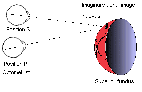
Whilst observing the aerial image of the fundus, the
practitioner should imagine that the fundus image is convex and not its true
concave form. In other words the practitioner should treat the image as if
looking at the convex surface of the moon (fig 1)

In figure 1 a naevus has been observed by the optometrist (position P) on
the inferior fundus (seen superiorly). The optometrist should move up
(position S) or ask the patient to look down to view the lesion.
With
direct ophthalmoscopy in mind, the optometrist would have been tempted to move
down or ask the patient to look up. In applying this method to record keeping,
and with the patient in a sitting position, the practitioner should make
his/her drawing with the record card upside down, and any later reference to
the fundus by direct ophthalmoscopy will be correctly orientated.
Summary
Indirect ophthalmoscopy gives rise to:
- an inverted image
- a laterally reversed image
The practitioner can reduce difficulties from the above when recording findings by turning the record card upside down whilst drawing the fundus appearance.
In addition difficulties in obtaining a view of different areas of the fundus may be helped by imagining a third change in perspective:- - an imaginary convex appearance to the fundus.